lymphoid follicles throat
1 Peyers patches are small clusters of lymphoid follicles found throughout the mucus membrane of the small intestine especially along the ileum. While this community is not a substitute for doctors advice and we cannot treat or diagnose we find being able to communicate with others who have IBD is invaluable as we navigate our struggles and celebrate.

Laryngeal Ectopic Tonsil As A Cause Of Dysphonia A Case Report Sciencedirect
The outer cortex is composed of follicles of B cells so that it is called the B-cell zone.
. They may sometimes even appear larger or more inflamed if you come down with a bad cold or the flu. These lymphoid tissues are controlled by specialized cells that arm themselves to attack and destroy foreign invaderssuch as bacteria fungi or virusesthrough phagocytosis or the production of antibodies. FLH in the oral and maxillofacial region is an uncommon benign entity which may resemble malignant lymphoma clinically and histologically.
I have enlarged lympiod follicles in my throat that were seen by an ent. They may contribute to the chronic. Primary follicles are lymphoid follicles that do not yet contain a germinal center described below.
25103 They are seen in 50 of barium studies performed on children and 13 of air-contrast barium enemas in adults. Welcome to Crohns Forum a support group for people with all forms of IBD. It is usually self-limiting and recovers naturally or with symptomatic treatment.
Nodular lymphoid hyperplasia of the gastrointestinal tract produces multiple discrete mucosal nodules in a variable segment of the small intestine large intestine or both. Lymphoid follicles appear as 1- to 3-mm elevations in the mucosa without a ring shadow. Follicular lymphoma is a non-Hodgkins lymphoma.
Infected Isolated Pharyngeal Lymphoids Inflamed Scattered Lymphoid Follicles of Posterior Pharyngeal Wall. 2 These are an important part of gut associated lymphoid tissue. Similar to a lymphoid aggregate sometimes used interchangeably but typically refers to a more discrete collection of B cells T cells and supporting cells.
The ENT examined me and looked at the lumps through my nose with a Flexible Laryngoscopy. As we grasp a better. They are also called aggregated lymphoid nodules.
Their function is to drain the fluid from the throat and nose and tongue region so that your immune cells in these lymphoid follicles can screen for infection to help make the specific antibodies when necessary. Lymphoid follicles have an average macroscopic density of 38 per centimeter of adult human colon. Cutaneous lymphoid hyperplasia is generally not malignant but in rare.
He said they were lymphoid tissue and that they were like little tonsils that pop up during infections I had my tonsils removed as a child and he said he wasnt concerned about them as they were normal anatomy. Citation needed Cutaneous hyperplasia Cutaneous lymphoid lesions may be observed in follicular granulomatous or lymphoreticular pathologic patterns. Aggregates of lymphoid tissue are all over the oral mucosa but they are often prominent in the soft palate uvula and pharynx.
An ectopic tonsil is tonsillar tissue that was. However their importance in COPD is unproven and their roles in pathogenesis are largely unknown. There are two types of lymphoid follicle.
Crohns Disease Forum. We observed histological features of hyperplastic tonsil for example existence of germinal center lymphoid tissue and crypt involving lymphoepithelial symbiosis Figure Figure2. Similarly the inner cortex has T cells and is called the T-cell zone.
On 18 September 2008 the tissue was resected laryngoscopically under general anaesthesia and this sample was pathologically analyzed. Here we propose that secondary and tertiary lymphoid tissues are part of a spectrum of lymphoid tissues that ranges from highly organized encapsulated tissues which develop at specific sites according to pre-programmed developmental pathways to granulomas and ectopic follicles which develop only after local inflammation or infection at a variety of sites in multiple tissues. We report the case of a 51-year-old woman who presented with an.
Lymphoid follicles LFs can be induced in the lung on infection or chronic inflammation. He said the rest looks normal its just the back of my throat but it like having a persistent sore throat. In some cases the etiology of FLH is unclear.
However their relevance and contribution to protective immunity or pathogenesis is poorly understood. When you have follicular lymphoma the sick blood cells can travel to many parts of your body such as. The numbers of lymphoid follicles are increased in clinical and experimental COPD 8 9.
Follicular lymphoid hyperplasia FLH is characterized by an increased number and size of lymphoid follicles. The lymphoid infiltrate is confined to the lamina propria and superficial submucosa. Nodular lymphoid hyperplasia is most commonly encountered.
Find high-quality stock photos that you wont find anywhere else. What does lymphoid follicles mean. Lymphoid follicles are small masses of tissues that contain aggregations of inflammatory cells mainly B cells with some T cells and dendritic cells.
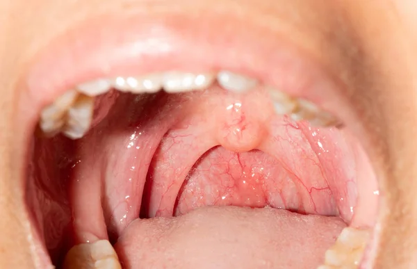
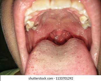
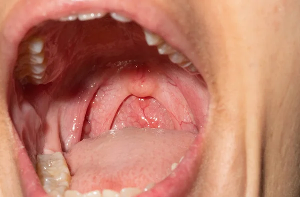
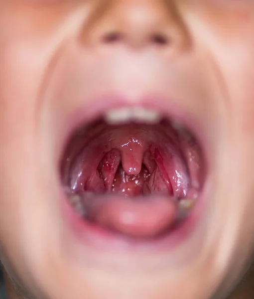
Search from Lymphoid Follicles stock photos pictures and royalty-free images from iStock. An injected throat caused by infective inflammation involving pharyngeal mucous membranes and lymphoid follicles. The lymphoid follicles of the throat are swollen and often covered with white exudate and cervical lymph nodes are enlarged and tender Pharyngeal Erythema Definition Pharyngeal relates to the pharynx part of your throatoral cavity.
Gastric involvement is rare. You dont need to worry about them. On the 6th September I saw an ENT as I had bumps in my throat.
Recent advances from clinical studies and animal models have shed some light on the mechanisms that trigger and facilitate the development of LFs.

Chronic Pharyngitis Causes Symptoms And Treatment Medchrome

Reply To Influenza Follicles And Their Buds As Early Diagnostic Markers Of Influenza Typical Images And Demonstration Of Lymphoid Follicles In The Posterior Pharyngeal Walls Of Patients With Mycoplasmal Pneumonia Postgraduate
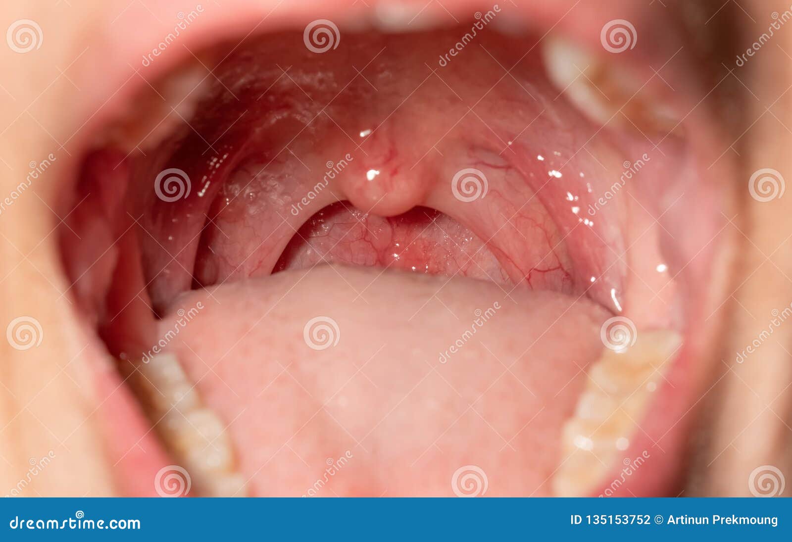
Sore Throat With Throat Swollen Closeup Open Mouth With Posterior Pharyngeal Wall Swelling And Uvula And Tonsil Influenza Stock Photo Image Of Background Follicles 135153752

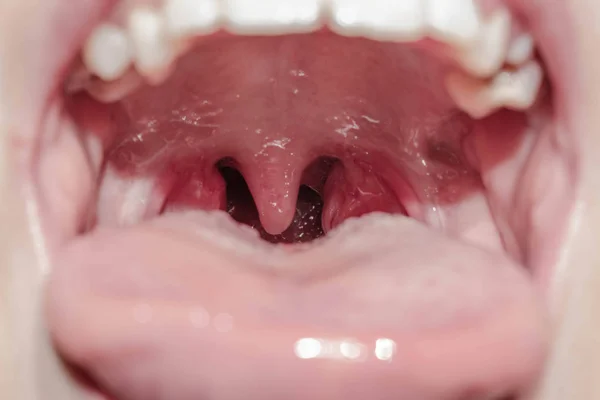
Tonsil Pharynx Otorhinolaryngology Portal
Pharynx Stock Photos Royalty Free Pharynx Images Depositphotos
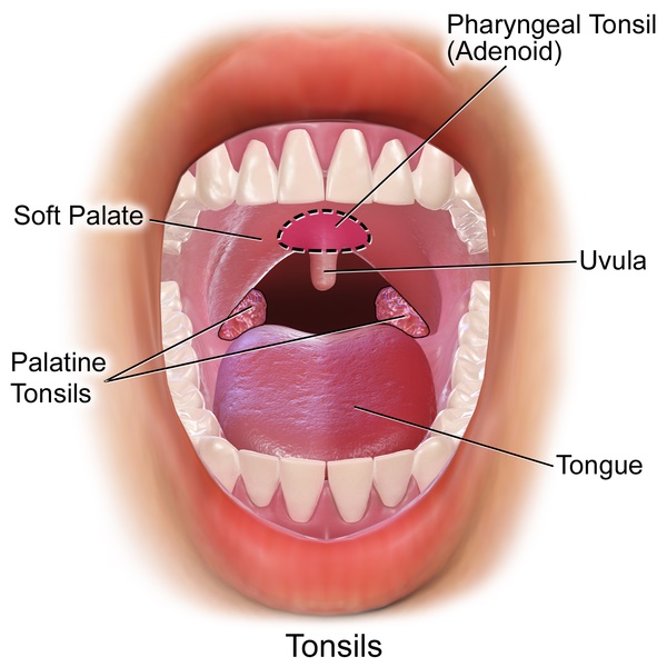
What Is The Difference Between Tonsils And Lymph Nodes Pediaa Com
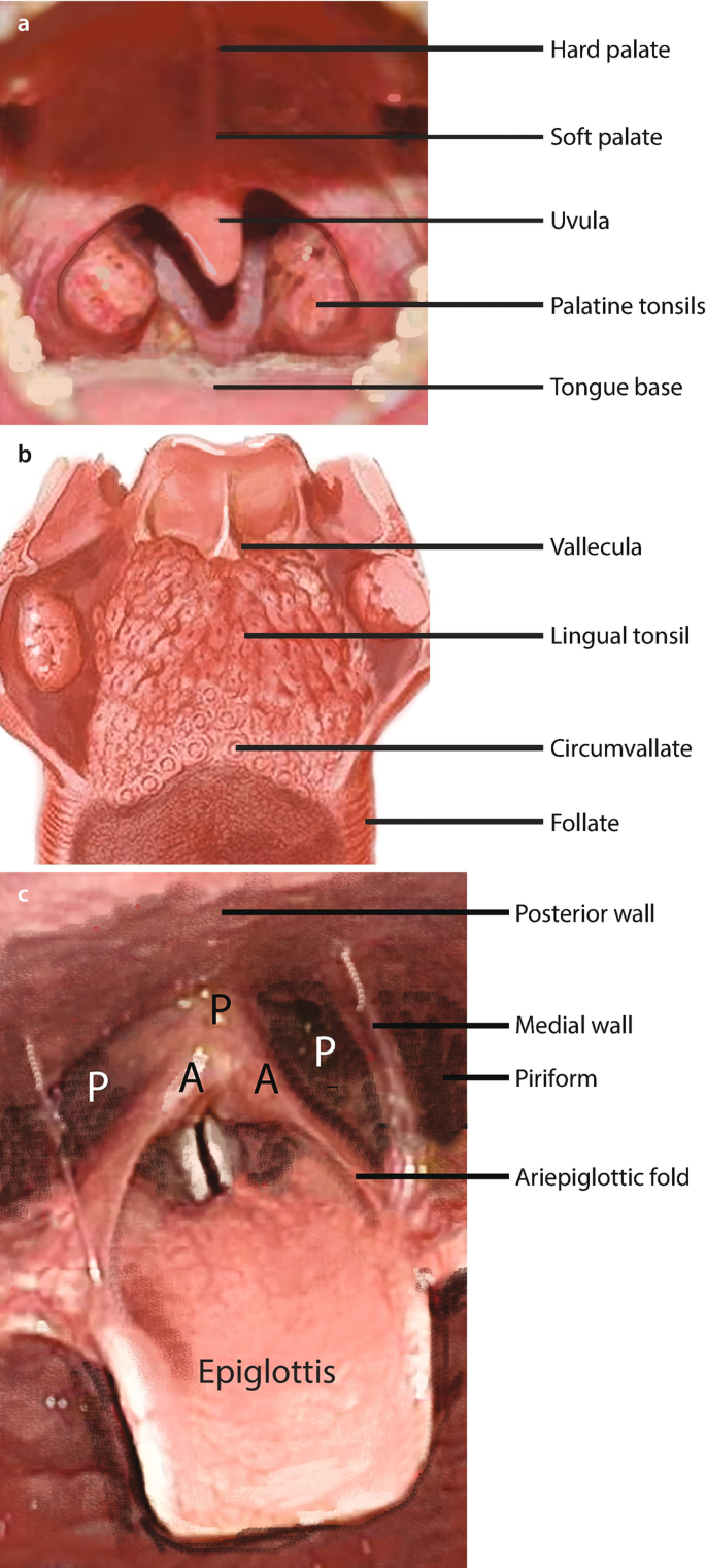
Oropharynx Hypopharynx And Parapharyngeal Space Anatomy Histology Benign And Malignant Neoplasia Springerlink
Lymphoid Stock Photos Royalty Free Lymphoid Images Depositphotos

Useful Clinical Findings And Simple Laboratory Data For The Diagnosis Of Seasonal Influenza Takeoka 2021 Journal Of General And Family Medicine Wiley Online Library
Similar Images Stock Photos Vectors Of Sore Throat With Throat Swollen Closeup Open Mouth With Posterior Pharyngeal Wall Swelling And Uvula And Tonsil Influenza Follicles In The Posterior Pharyngeal Wall Macro

Sore Throat Throat Swollen Closeup Open Stock Photo 1319726006 Shutterstock
Lymphoid Stock Photos Royalty Free Lymphoid Images Depositphotos
Tonsil Stock Photos Royalty Free Tonsil Images Depositphotos

Free Art Print Of Sore Throat With Throat Swollen Closeup Open Mouth With Posterior Pharyngeal Wall Swelling And Uvula And Tonsil Influenza Follicles In The Posterior Pharyngeal Wall Upper Respiratory Tract

Pharyngeal Findings From Case 2 Lymphoid Follicles With Diameters Of 5 Download Scientific Diagram
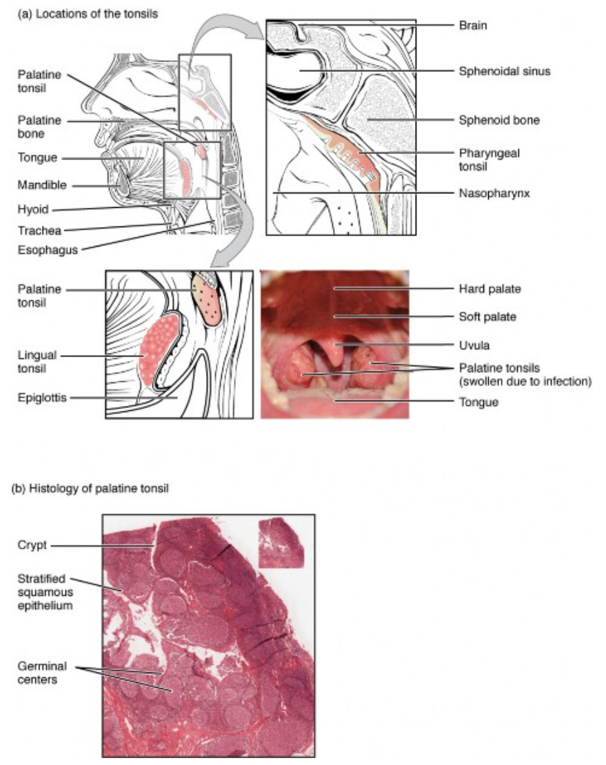
18 5 Lymphoid Nodules Biology Libretexts

7 023 Soft Palate Stock Photos Pictures Royalty Free Images Istock

Sore Throat With Throat Swollen Closeup Open Mouth With Posterior Pharyngeal Wall Swelling And Uvula And Tonsil Influenza Follicles In The Posterior Stock Photo Alamy

A A Very Rare Case Where Influenza Follicles And Influenza Follicle Download Scientific Diagram


Comments
Post a Comment